
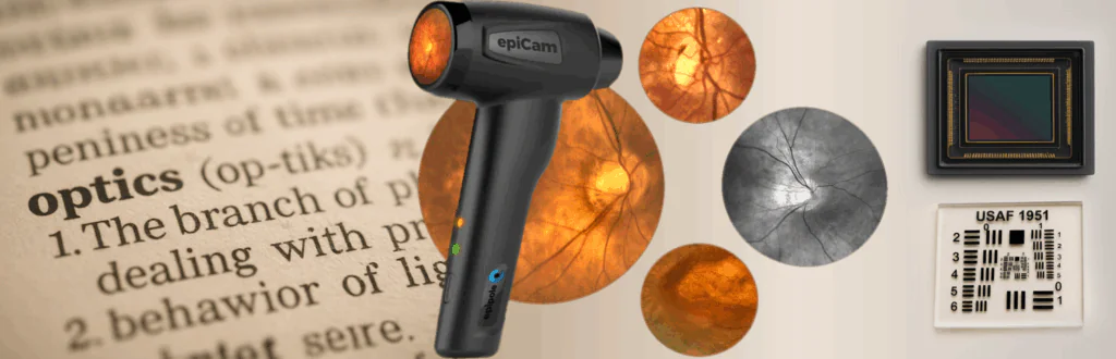
Dr Ian Murray, R&D, epipole
September 2025
Introduction
Two key concepts influencing fundus camera performance are pixel resolution and optical resolution. While they can sometimes be conflated, these terms represent distinct aspects of imaging technology. Pixel resolution refers to the number of pixels in the digital image captured by the camera, while optical resolution refers to the inherent ability of the camera’s lens system to distinguish fine details. This paper aims to clarify the differences between pixel and optical resolution, discuss their interplay, and explore their implications for fundus cameras, with specific reference to epipole’s own fundus camera – the epiCam®
When evaluating digital fundus cameras, it is easy to assume that a higher pixel count directly translates to better image quality. Many manufacturers, mirroring trends in consumer digital cameras, highlight megapixel (MP) counts as a key indicator of performance. While pixel resolution is an important aspect of imaging, it is not the sole determinant of a fundus camera’s ability to capture clinically useful detail.
Pixel Resolution
Pixel resolution refers to the number of pixels that make up a digital image. It represents the image’s dimensions in terms of pixel count, usually expressed as width by height, and often the total number of pixels, in megapixels (MP). The pixel resolution of an image is determined by the imaging sensor of the camera, which is composed of an array of photodetectors that capture light and convert it into electrical signals. A higher pixel resolution over the same physical area can correspond to greater detail in the image, as each pixel represents a smaller portion of the overall scene being captured (figure 1).
However, while pixel resolution is a factor in determining the level of detail in a digital image, the optical lens system in front of the image sensor plays an extremely important role in the resolution of the camera. The lens system itself also has a resolution, known as its optical resolution.
Figure 1. Illustration of pixel resolution differences between a 5 MP sensor (2592 × 1944 pixels) and a 12 MP sensor (4000 × 3000 pixels) over the same physical area. The magnified insets highlight that the higher resolution sensor has a greater number of smaller pixels.
Optical Resolution
Optical resolution is a measure of the ability of a lens system to resolve fine details in an object being imaged. It is determined by the quality and configuration of the camera’s optical components. Unlike pixel resolution, which is defined by the image sensor, optical resolution depends on the camera’s ability to focus light accurately onto the sensor.
Optical resolution is often expressed in terms of line pairs per millimeter (lp/mm), which indicates the smallest distance between two lines that the optical system can distinguish as separate. A better optical resolution enables the system to resolve finer details (figure 2). In fundus cameras, this is essential for capturing clinically relevant features such as small blood vessels and microaneurysms in the retina. Even if the image sensor has a high pixel resolution, poor optical resolution can result in blurry or indistinct images, rendering the additional pixels ineffective. Conversely, an optical system with excellent optical resolving power can capture detailed images even with a moderate pixel count, provided the sensor’s pixel size is sufficiently small to match the optical resolution.
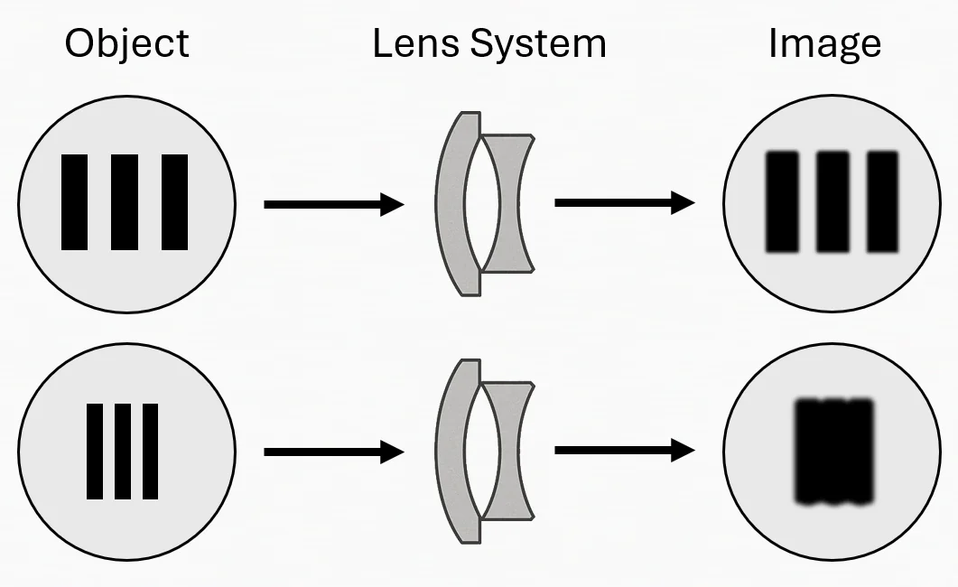
Figure 2. Illustration of optical resolution of a lens system. On the the top, an object containing a set of parallel black line pairs can be resolved (distinguished) when imaged by the optical lens system system. The bottom demonstrates imaging of higher-frequency details (closely spaced lines) which become indistinguishable by this lens system, illustrating that the optical resolution limit of the lens system has been passed.
Relationship Between Pixel Resolution and Optical Resolution
Pixel resolution and optical resolution are closely interconnected, but they are not interchangeable. While pixel resolution defines the number of discrete points used to construct a digital image, optical resolution determines how much detail the optical system can deliver to the sensor. The interplay between these two factors dictates the overall quality of the image.
Increased pixel resolution over the same area means more numerous pixels that are smaller. Smaller pixels allow the sensor to capture more detailed images, as they correspond to smaller portions of the image projected by the optical system. However, this benefit is only realised if the optical resolution of the system is high enough to resolve details at the scale of the pixel size.
If the optical resolution exceeds the resolving power of the sensor, details that could have been captured by the optics are lost due to the insufficient pixel density of the sensor. This phenomenon, known as under-sampling, results in images that lack the clarity and detail that the optical system is capable of providing. On the other hand, if the pixel size is too small relative to the optical resolution, the system is said to be over-sampling. While over-sampling can theoretically capture more information, it can also lead to unnecessary additional problems without a corresponding improvement in resolving capability of the camera. For example, smaller pixels can mean lower sensitivity to light and higher noise levels in low light environments because the smaller photodetectors capture less light. This trade-off between pixel resolution and optical resolution is critical in fundus imaging, where the ability to visualise fine retinal structures can have significant clinical implications. Larger pixels, while less capable of capturing fine details, generally offer better light sensitivity, which is advantageous for imaging in dim conditions or when capturing subtle retinal features. If the pixel size is too large, these critical details may be missed. Conversely, excessively small pixels may result in images with high noise levels, especially in low-light conditions, which may also obscure important features.
An optimal imaging system matches the pixel resolution of the sensor to the optical resolution of the lens system. This ensures that the details resolved by the optics are captured accurately by the sensor, without unnecessary over-sampling or under-sampling. For fundus cameras, achieving this balance requires careful consideration of both the optical design and the sensor specifications. Systems that prioritise one aspect at the expense of the other will produce images that are suboptimal.
epipole’s fundus camera, epiCam®, has been designed to optimally balance its sensor pixel resolution and the optical resolution of the epiCam®lens system, while also taking into account many other factors (such as the camera field of view, video frame rate and data bandwidth aspects for real-time HD video encoding). The epiCam® uses high quality optics and a lens system designed to meet and exceed the international standard ISO 10940 (“Ophthalmic instruments – Fundus cameras”) which specifies image quality requirements and test methods for fundus cameras, including optical resolution requirements.
Why it matters: Practical Implications for Fundus Cameras
In fundus imaging, high pixel counts often grab attention, but good optical resolution is essential for capturing high-quality, clinically useful images. High pixel resolution can enhance the detail captured in an image, but only if the optical system is capable of resolving those details. Cameras with excellent optical resolution will be better equipped to identify subtle pathological changes, such as microaneurysms and early retinal neovascularization. To provide further context to this, Table 1 lists optical resolution in line pairs per mm, and µm (meaning the smallest resolvable object size) alongside retinal pathology examples of approximately equivalent size. epipole’s epiCam® meets ISO 10940 which demands an optical resolution of 60 lp/mm in the image centre.
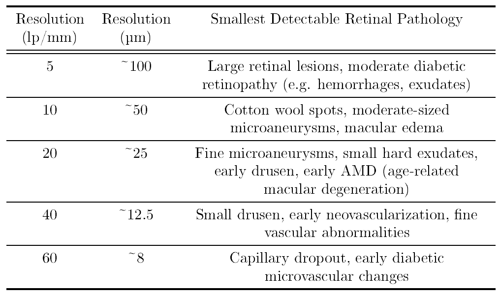
Table 1. Optical resolution and detectable retinal pathologies. Optical resolution expressed in line pairs per millimeter (lp/mm) and micrometers (µm), alongside approximate examples of retinal pathologies that can be detected at each resolution.
While marketing materials can sometimes emphasise the megapixel count of a camera, this metric alone does not guarantee superior imaging quality and when selecting a fundus camera, it is essential to evaluate the optical resolution to gain a meaningful understanding of its capabilities. High-pixel sensors may increase the cost of the camera without providing significant clinical benefits unless paired with high-quality optics. In some cases, systems with moderate pixel resolutions but excellent optical performance may be more cost-effective and clinically useful. Ultimately, the best imaging systems integrate high-quality optics with sensors that maximally utilise the resolving power of the optical system, providing clear, detailed retinal images for effective diagnosis and management of ocular diseases.
–
Download this white paper here
We love to talk!
Drop us a line at https://www.epipole.com/contact/