
Introducing HD Video Recording Capabilities in Retinal Imaging
In a groundbreaking leap forward for the field of mobile optometry, we are thrilled to announce the latest advancement in retinal imaging technology in our epiCam product: the integration of high-definition (HD) video capabilities across three different imaging modalities, and a user interface that is easier to use than ever:
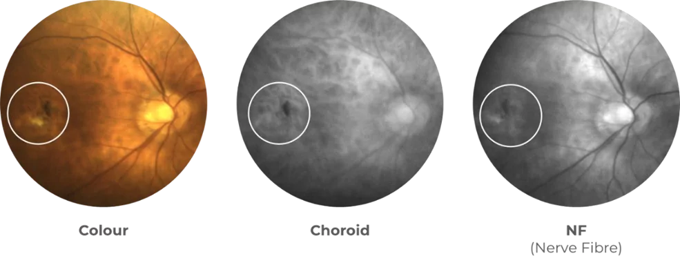
Example imaging from full color, Choroidal (Deep Red) and Nerve Fibre (Amber) Illuminants
Changing the Industry
The integration of multi-illuminant HD video functionality into retinal imaging devices signifies a transformative shift in the way optometrists can visualize and analyze ocular health – especially related to mobile optometry and out of practice care.
Traditionally, retinal/fundus imaging has primarily relied on static images to assess the condition of the retina. These are all still available with the epiCam of course, but with the introduction of HD video capabilities, especially in such a portable device, optometrists can now capture real-time footage of the retina in three distinct video imaging modalities: Full Color, Nerve Fibre (Amber) and Choroidal (Deep Red), all in a variety of locations, wherever they need to conduct examinations.
This means that not only can optometrists obtain high-resolution images of the retina, but they can also observe and capture things like the movement of retinal blood vessels (e.g. spontaneous venous pulse). epiCam retinal video recording has been used already in some fascinating research, linking observations in the retina with wider health issues (full details available upon request) and we’re excited to see the additional applications as they develop.
See More, Share More
Alongside the technical functionality improvements, there have been comprehensive developments in the software supporting the imaging capture too, enabling optometrists (and medical professionals) to easily compare imagery and video from previous imaging sessions, and to quickly and clearly monitor changes over time, accurately visualizing over different layers of the retina.
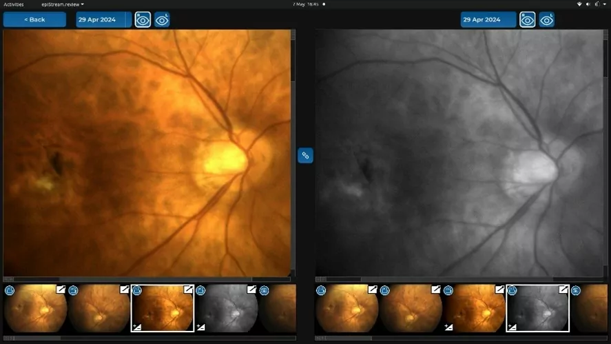
Compare and contrast – different sessions, different layers, same device
Expertly Developed
Dr Ian Murray, Head of R&D at epipole, sat down with the team and spoke excitedly about the latest iteration of our award-winning camera:
“We’ve been working with our partners and customers over the past year, incorporating all the feedback we’ve received, to ensure our ‘real world’ users are at the core of our product development, and I have to say we’re really pleased with the results.
We now have an updated, more user-friendly interface, where epiCam users can now select both ‘Advanced’ and ‘Guided’ modes, helping make it easier than ever for more people to use.
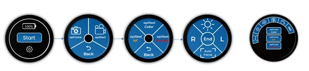
More power to the users – Guided and Advanced Mode screenshots, making imaging more accessible than ever before
Available in both modes, users can now record entire retinal examinations with the different illuminants (Amber/White Light/Deep Red), all on a single device.
This has all been done to support access by a wider range of practitioners to examine and record different views of the retina (full color imaging with white light, a view of the deeper retina with red, and the anterior to mid-retina with amber) with just a click of a button or a tap on a screen, and has been designed with the intent to further promote efficiency in clinical settings, and we’ve documented examples of how this can help improve patient flow, and optimize practice productivity.
There’s the wider public health angle we’re really proud of too – By offering patients the opportunity to actually view their examinations like never before, actually being able to see their retina in such high quality video, we’re hoping to help empower them to take a more active role in their eye health journey with the doctors being able to show them exactly what they are seeing.”
More Flexibility than Ever Before
As Dr Murray says, epiCam users can now capture entire examinations from start to finish in HD video, with three (3) imaging modes/illuminants.
To help simplify the operational nomenclature, this retinal video footage is called an epiReel, and is an additional option alongside the epiBurst image capture functionality (which operates similarly to a ‘live Photo’ burst photo on the iphone) which captures a series of individual frames in one go.
The users can review and select the exact epiFrames (individual images/frames) from these epiReels and epiBursts, for billing purposes, as well as for further examination, comparison, reporting, annotation and/or sharing.
Embedded video showing an example color (white light) retinal imaging session
Mobile First
One of the most significant implications of this advancement is its potential to revolutionize mobile retinal imaging, particularly for out-of-practice settings.
Traditionally, accessing specialized retinal imaging equipment has been limited to clinical settings, requiring patients to visit optometry offices or hospitals for evaluation. However, with the improvements to the ease of use making HD video capture easier than ever before, eye care professionals of all levels of experience now have access to a device that enables them to bring advanced retinal imaging directly to their patients; whether they are in remote locations, nursing homes, corporate setting, or community health centers (to name but a few potential locations!)
This ability to perform mobile retinal imaging has the potential to be a game changer for the optometry industry in the United States, and around the world.
By eliminating the need for patients to travel to specialized facilities for retinal evaluations, optometrists can enhance access to care, particularly for underserved populations with limited mobility or geographic barriers.
Additionally, the real-time visualization provided by HD video capabilities enables optometrists to make more informed clinical decisions on the spot, potentially leading to earlier detection and intervention for sight-threatening conditions – an extension of our commitment to our mission of the ‘eradication of preventable blindness.’
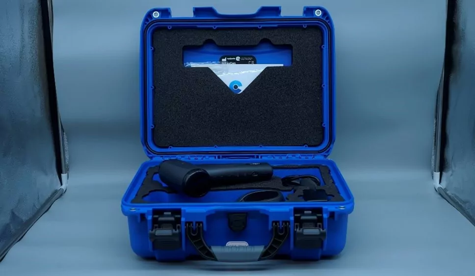
Working towards our goal of “Wherever there is an eye exam, there is an epiCam” by providing the very latest in ultraportable imaging solutions to eyecare professionals around the world
The Future of Mobile Retinal Imaging is Here
In conclusion, the integration of HD video capabilities into retinal imaging represents a groundbreaking advancement that has the power to elevate the standard of care in remote optometry.
By providing optometrists with the ability to capture real-time, high-definition footage of the retina in three different imaging modalities, this innovative device opens up new possibilities for imaging, examinations, and patient engagement, and we can’t wait to see what our partners do with the technology!
To find out how an epiCam can help you ‘See More’, get in touch to arrange a demonstration: https://www.epipole.com/arrange-a-demo/